Art-labeling activity neuroglial cells of the cns – Art-labeling activity of neuroglial cells in the CNS unveils a captivating realm of neuroscience, inviting us to explore the intricacies of these enigmatic cells. This technique, like a celestial cartographer, charts the vast expanse of neuroglial functions and dysfunctions, guiding us toward a deeper understanding of their role in neurological health and disease.
Neuroglial cells, the unsung heroes of the nervous system, have long been overshadowed by their neuronal counterparts. However, recent advances in art-labeling techniques have thrust these cells into the spotlight, revealing their remarkable diversity and essential contributions to neural function.
Introduction: Art-labeling Activity Neuroglial Cells Of The Cns

Art-labeling activity is a powerful technique for studying the neuroglial cells of the central nervous system (CNS). By labeling neuroglial cells with fluorescent or other markers, researchers can visualize and track these cells in living animals or in tissue culture.
This technique has provided important insights into the development, function, and dysfunction of neuroglial cells.
Neuroglial cells are a diverse group of cells that make up the supporting tissue of the CNS. They outnumber neurons by 10 to 1 and play a critical role in maintaining the health and function of the nervous system. Neuroglial cells provide structural support, regulate the extracellular environment, and modulate neuronal activity.
They also play a role in neurogenesis, synaptogenesis, and immune responses in the CNS.
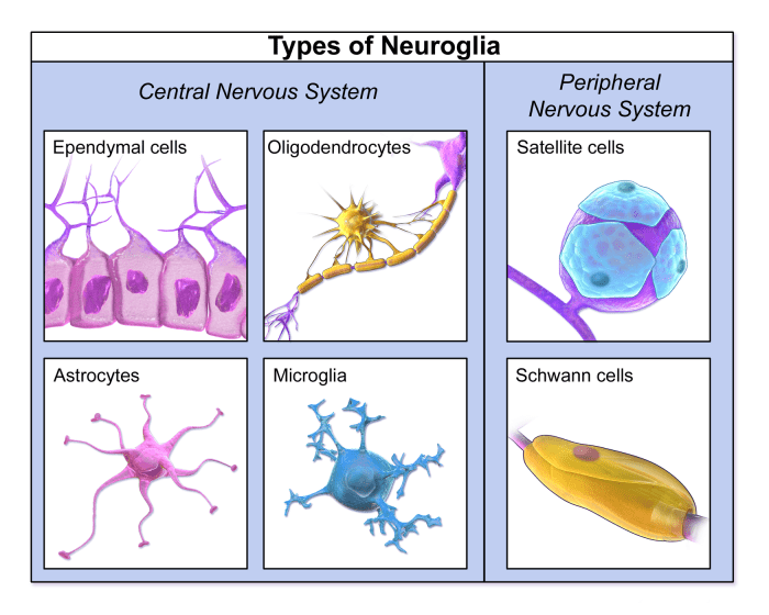
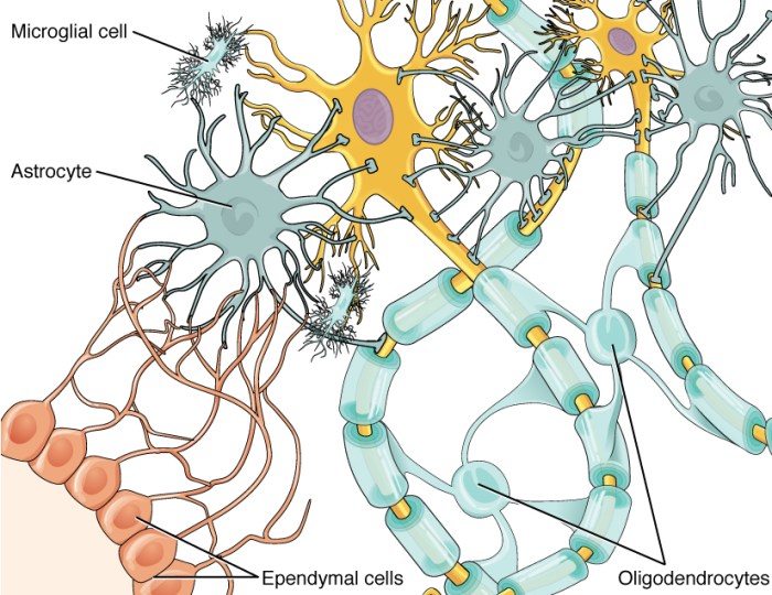
Art-labeling activity has been used to study a wide range of neuroglial cell types, including astrocytes, oligodendrocytes, microglia, and ependymal cells. These studies have provided insights into the development, function, and dysfunction of these cells in both normal and pathological conditions.
Methods and Procedures

There are a variety of methods that can be used to art-label neuroglial cells. The most common method is immunohistochemistry, which involves using antibodies to label specific proteins expressed by neuroglial cells. Other methods include genetic labeling, which involves introducing a fluorescent or other marker gene into neuroglial cells, and viral labeling, which involves using a virus to deliver a fluorescent or other marker gene to neuroglial cells.
Each method has its own advantages and disadvantages. Immunohistochemistry is a relatively simple and inexpensive method, but it can only be used to label proteins that are expressed by neuroglial cells. Genetic labeling is a more permanent method, but it can be more difficult and expensive to introduce a fluorescent or other marker gene into neuroglial cells.
Viral labeling is a very efficient method, but it can be more difficult to control the expression of the fluorescent or other marker gene.
The following is a detailed protocol for immunohistochemistry labeling of neuroglial cells:
- Perfuse the animal with 4% paraformaldehyde in phosphate-buffered saline (PBS).
- Post-fix the tissue in 4% paraformaldehyde in PBS for 24 hours.
- Cryosection the tissue into 10-μm-thick sections.
- Block the sections in 10% normal goat serum in PBS for 1 hour.
- Incubate the sections with the primary antibody in 10% normal goat serum in PBS for 24 hours.
- Wash the sections with PBS for 3 × 10 minutes.
- Incubate the sections with the secondary antibody in 10% normal goat serum in PBS for 1 hour.
- Wash the sections with PBS for 3 × 10 minutes.
- Mount the sections on slides and coverslip them.
Results

Art-labeling studies have provided important insights into the development, function, and dysfunction of neuroglial cells. These studies have shown that neuroglial cells are highly dynamic cells that play a critical role in the health and function of the nervous system.
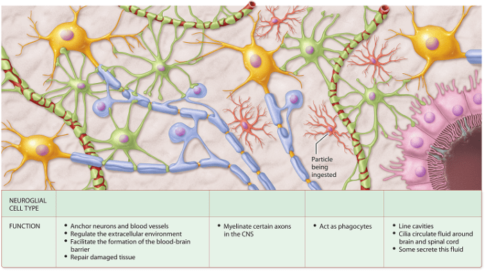
For example, art-labeling studies have shown that astrocytes are involved in the formation of the blood-brain barrier, the regulation of extracellular ion concentrations, and the release of neurotransmitters. Oligodendrocytes are involved in the formation and maintenance of myelin, which insulates axons and speeds up the transmission of electrical signals.
Microglia are involved in the immune response of the CNS, and they can phagocytose damaged neurons and other cells.
Art-labeling studies have also provided insights into the role of neuroglial cells in neurological disorders. For example, studies have shown that astrocytes and microglia are activated in response to brain injury, and that this activation can contribute to the development of secondary damage.
Oligodendrocytes are damaged in multiple sclerosis, and this damage can lead to demyelination and axonal loss.
Discussion
Art-labeling activity is a powerful technique for studying the neuroglial cells of the CNS. This technique has provided important insights into the development, function, and dysfunction of these cells. However, there are still many unanswered questions about the role of neuroglial cells in the nervous system.
Future research using art-labeling activity will help to answer these questions and provide a better understanding of the role of neuroglial cells in health and disease.
Applications

Art-labeling activity has a wide range of applications in the study of the neuroglial cells of the CNS. This technique can be used to study the development, function, and dysfunction of these cells in both normal and pathological conditions.
Art-labeling activity can also be used to develop new treatments for neurological disorders. For example, art-labeling activity has been used to develop new drugs that target astrocytes and microglia. These drugs have shown promise in animal models of neurological disorders, and they may eventually be used to treat patients with these disorders.
Question & Answer Hub
What is art-labeling activity?
Art-labeling activity refers to a range of techniques used to visualize and study neuroglial cells in the CNS. These techniques involve introducing a tracer or marker into the cells, allowing researchers to track their morphology, connectivity, and function.
Why is studying neuroglial cells important?
Neuroglial cells play crucial roles in supporting neuronal function, maintaining homeostasis, and mediating immune responses in the CNS. Understanding their activity is essential for deciphering the complex interplay between neurons and glia in health and disease.
What are the applications of art-labeling activity in neuroglial research?
Art-labeling activity has wide-ranging applications in neuroglial research, including mapping neural circuits, investigating cellular interactions, and identifying molecular markers associated with neuroglial dysfunction.